International Association for Integrated Oncology (IAIO) takes pride in its commitment to providing a platform to clinicians, practitioners, and cancer professionals for sharing their knowledge through continuous stream of medical education programs. IAIO serves as a collaborative hub for cancer science experts, practitioners, and influential professionals in the oncology field. Our primary objective has been the establishment of scientific communities and special interest groups, all in the pursuit of disseminating knowledge and facilitating education. Our goal is to provide online and in-person continuing medical education (CME) on oncology and its related emerging clinical topics that will improve physician practice and patient care.
View More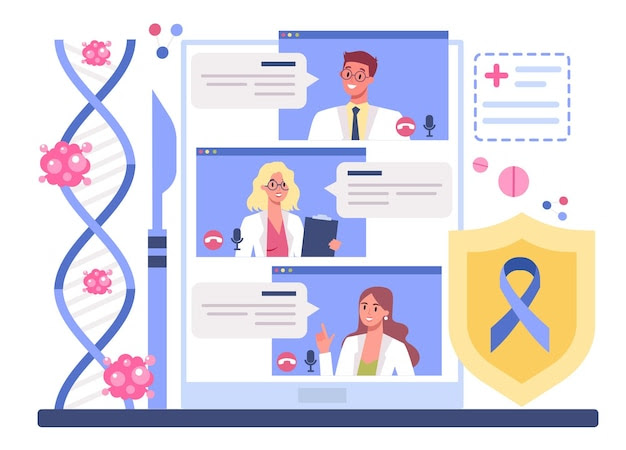
The IAIO, as one of the significant professional associations, strives to gather the researchers, academicians, and students in a single forum by educating young and experienced specialists through scientific sessions on current victories in medical oncology & life science.
Apply NowWe invite you to be a part of this prestigious event as a sponsor or exhibitor. Our goal is to provide an engaging and informative platform for you to showcase your products and services, network with professionals in the field and contribute to the advancement of n Clinical and Medical oncology.
Apply Now
Cancer nanomedicine technology is a field of medicine that utilizes nanoscale particles to diagnose and treat cancer. These nanoscale particles are engineered to target specific cells and tissues in order to deliver drugs and other therapies directly to the tumour site.

Advanced cancer that has spread beyond the initial tumour to other parts of the body. Treatment for advanced cancer depends on the type of cancer, the location of the cancer, and the patient's overall health.
An Advanced Clinical Practitioner Oncology is an advanced practice nurse, physician assistant, certified registered nurse anaesthetist, or other healthcare provider who specializes in the care of cancer patients.
Radiology technology is the medical specialty of using imaging techniques such as X-rays, Computed Tomography (CT), Magnetic Resonance Imaging (MRI), nuclear medicine and ultrasound to diagnose and treat medical conditions.
All the Abstracts/Full Paper for the Upcoming conferences can be submitted here.
All the research articles for publication in Journals SCOPUS Indexed can be submitted here.
For any kind of research assistance please fill this form (redirect to the form in research assistance)